Search Result
Results for "
fluorescent stain
" in MedChemExpress (MCE) Product Catalog:
4
Biochemical Assay Reagents
| Cat. No. |
Product Name |
Target |
Research Areas |
Chemical Structure |
-
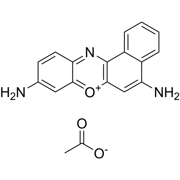
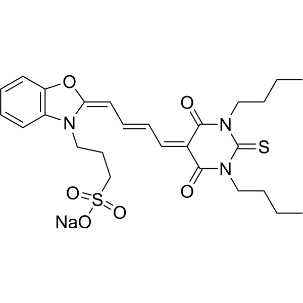
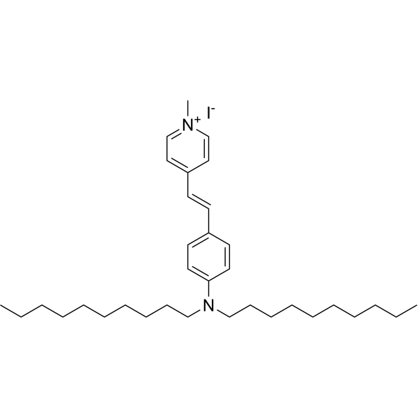
- HY-101888
-
-
-
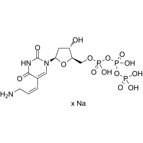
- HY-D1674
-
|
|
Fluorescent Dye
|
Others
|
|
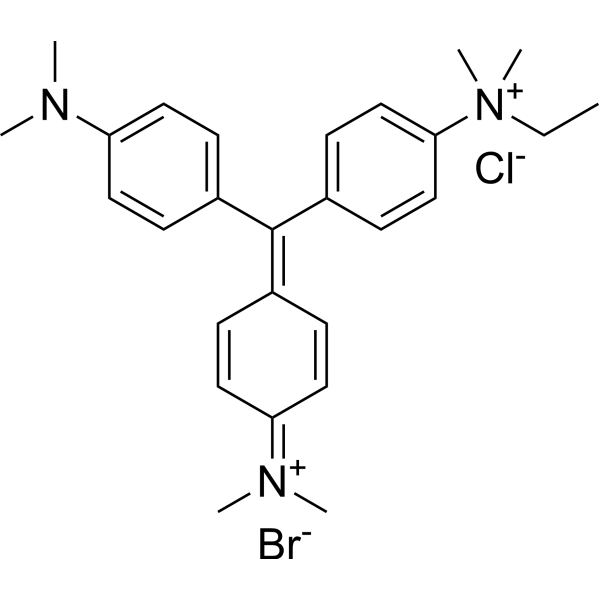
Sulforhodamine G is a fluorescent stain with broad dynamic ranges. Sulforhodamine G can be used for the research of protein stains .
|
-
-
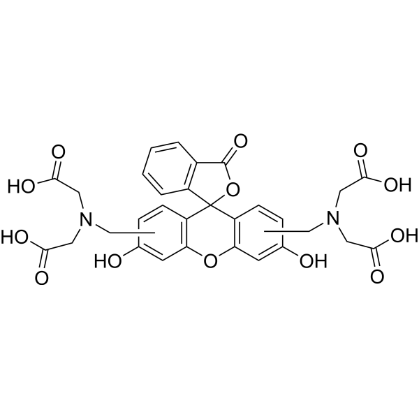
- HY-101889
-
-
-
- HY-D1021
-
|
Aminoallyl-dUTP sodium salt
|
DNA/RNA Synthesis
|
Others
|
|
AA-dUTP sodium salt is a fluorescent dye which can be used to stain cDNA.
|
-
-
- HY-107629
-
-
-
- HY-Y0016
-
|
Basic Violet 10; Brilliant Pink B; Rhodamine O; Tetraethylrhodamine
|
Fluorescent Dye
|
Others
|
|
Rhodamine B is a staining fluorescent dye, commonly used for dyeing textiles, paper, soap, leather, and agents.
|
-
-
- HY-D1775
-
|
|
Fluorescent Dye
|
Others
|
|
Lysotracker blue DND-22 is a blue-fluorescent probe for staining acidic compartments in live cells.
|
-
-
- HY-117070
-
|
|
Fluorescent Dye
|
Others
|
|
TO-PRO-3 iodide is a highly efficient blue fluorescent dye that can stain cytoplasm as a cell tracer.
|
-
-
- HY-D0163
-
|
|
DNA Stain
|
Others
|
|
Methyl Green is a potent fluorescent dye. Methyl Green is a DNA stains of cells and electrophoretic gels. Methyl Green can be used as a stain for direct measuring of viability by both microscopy and flow cytometry, with peaks at 633 and 677 nm .
|
-
-
- HY-D0021
-
|
EtBr; Homidium bromide
|
DNA Stain
|
Others
|
|
Ethidium bromide is an intercalating agent commonly used as a fluorescent tag (nucleic acid stain) in molecular biology laboratories for techniques such as agarose gel electrophoresis.
|
-
-
- HY-114227
-
|
|
DNA Stain
|
Inflammation/Immunology
|
|
Hexidium iodide, a fluorescent nucleic binding acid stain (excitation/emission ~ 518/600 nm), permeants to mammalian cells and selectively stains almost all gram-positive bacteria. Hexidium iodide can bind to the DNA of all bacteria after permeabilization by EDTA .
|
-
-
- HY-D0933
-
|
|
|
|
|
Auramine O is a yellow fluorescent dye and can be used to stain acid-fast bacteria. Auramine O is toxic and resistant in the environment .
|
-
-
- HY-D1475
-
|
|
Fluorescent Dye
|
Others
|
|
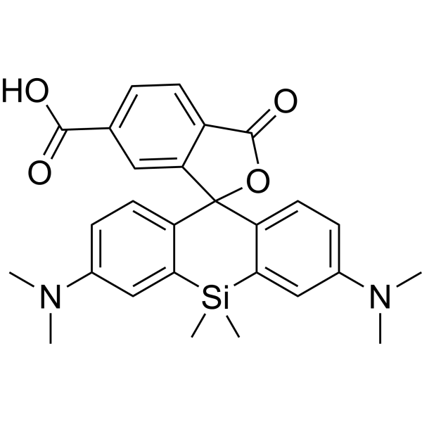
SIR-6-COOH is a fluorescent dye. SIR-6-COOH can be used for staining proteins in live-cell STED imaging
|
-
-
- HY-108166
-
|
|
Fluorescent Dye
|
Inflammation/Immunology
|
|
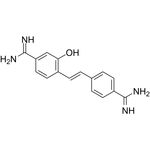
Hydroxystilbamidine, a dye capable of binding to both DNA and RNA, is a powerful inhibitor of cellular ribonucleases. Hydroxystilbamidine is a retrograde fluorescent tracer and a histochemical stain [1]
|
-
-
- HY-D1385
-
|
|
Fluorescent Dye
|
Others
|
|
JF526–Pepstatin A TFA is a fluorescent dye that can be used for lysosomal staining in live cells. The excitation maximum is 530 nm and the emission maximum is 549 nm .
|
-
-
- HY-D0718
-
|
Nile Blue A oxazone; Phenoxazone 9
|
Fluorescent Dye
|
Others
|
|
Nile red (Nile blue oxazone) is a lipophilic stain. Nile red has environment-sensitive fluorescence. Nile red is intensely fluorescent in a lipid-rich environment while it has minimal fluorescence in aqueous media. Nile red is an excellent vital stain for the detection of intracellular lipid droplets by fluorescence microscopy and flow cytof uorometry. Nile red stains intracellular lipid droplets red. The fluorescence wavelength is 559/635 nm .
|
-
-
- HY-D0127
-
|
|
Fluorescent Dye
|
Others
|
|
Merocyanin 540 is a fluorescent membrane probe that selectively stains the membranes of a wide variety of electrically excitable cells, but not those of nonexcitable cells (Ex/Em: 540/580 nm) .
|
-
-
- HY-D1493
-
|
|
PKC
|
Others
|
|
FIM-1 is a fluorescent PKC (protein kinase C) probe that can be used for mitochondrial staining. FIM-1 inhibits PKC and acts as ATP-competitive catalytic site inhibitor .
|
-
-
- HY-137896
-
|
|
Fluorescent Dye
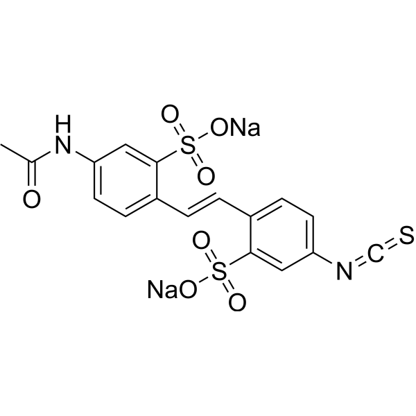
|
Others
|
|
4-Acetamido-4'-isothiocyanatostilbene-2,2'-disulfonic acid disodium is a fluorescent dye. 4-Acetamido-4'-isothiocyanatostilbene-2,2'-disulfonic acid disodium can be used to demonstrate retrograde axonal transport to label secondary antibodies and as a fluorescent whole cell stain .
|
-
-
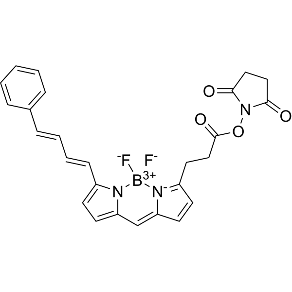
- HY-D1691
-
|
|
Fluorescent Dye
|
Others
|
|
BODIPY-581/591 NHS ester is a bright, red fluorescent dye (excitation: 581 nm; emission: 591 nm). BODIPY-581/591 NHS ester shows unique hydrophobic properties for staining lipids, membranes, and other lipophilic compounds .
|
-
-
- HY-D1249
-
|
|
Fluorescent Dye
|
Others
|
|
Calcein mixture of isomers is a calcium-dependent fluorescent molecule. Calcein mixture of isomers can be used to study bone metabolism (in vivo) and to stain depressed areas (in vitro). Calcein mixture of isomers can also be used for fluorometry and EDTA titration of calcium.
|
-
-
- HY-D1630
-
|
|
Fluorescent Dye
|
Others
|
|
4-Di-10-ASP is a fluorescent lipophilic tracer (Excitation 485 nm; Emission 620 nm). 4-Di-10-ASP can be used to stain phospholipid membranes in a specific manner .
|
-
-
- HY-D0895
-
-
-
- HY-D0950A
-
|
|
DNA Stain
|
Others
|
|
Methyl Green zinc chloride is a potent fluorescent dye. Methyl Green zinc chloride is a DNA stains of cells and electrophoretic gels. Methyl Green zinc chloride can be used as direct measuring of viability by both microscopy and flow cytometry, with peaks at 633 and 677 nm .
|
-
-
- HY-D1421
-
PKH 67
1 Publications Verification
|
Fluorescent Dye
|
Others
|
|
PKH67 is a fluorescent cell binding dye with green fluorescence. PKH67 can stain the cell membrane and the Ex/Em is 490/502 nm. PKH67 is often used in combination with the non-specific red fluorescent dye PKH26 (Ex/Em=551/567 nm) to label cells, detect cell proliferation in vitro, and trace cells in vitro and in vivo .
|
-

-
- HY-D1074
-
|
3,3'-Dipropyloxacarbocyanine iodide
|
Fluorescent Dye
|
Others
|
|
DiOC3(3) (3,3'-Dipropyloxacarbocyanine iodide) is a green fluorescent lipophilic dye with cell membrane permeability. DiOC3(3) can be used to stain cell membranes and other lipid-soluble biological structures .
|
-
-
- HY-D1020
-
-
-
- HY-135056
-
|
|
Fluorescent Dye
|
Others
|
|
Mito-Tracker Green is a green fluorescent dye that selectively accumulates in the mitochondrial matrix. MitoTracker Green FM covalently binds mitochondrial proteins by reacting with free mercaptan of cysteine residues, allowing staining of mitochondrial membrane potential independent of membrane potential. Excitation/emission wavelength 490/523 nm.
|
-
-
- HY-D1723
-
|
|
DNA Stain
|
Others
|
|
EthD-III is a nucleic acid probe. EthD-III is a red fluorescent stain that can be used to detect dead cells. EthD-III enters cells with damaged membranes and binds to nucleic acids, resulting in bright red fluorescence in dead cells (Ex/Em=530/645 nm) .
|
-
-
- HY-D0093
-
|
EthD-1
|
DNA Stain
|
Others
|
|
Ethidium homodimer (EthD-1) is a high-affinity fluorescent nucleic acid dye commonly used to stain mammals, bacteria, yeast, and fungi. Ethidium homodimer binds to DNA or RNA, enhancing fluorescence more than 30 times. The Ethidium homodimer has a strong positive charge, so it cannot cross cell membranes and stain living cells; But the Ethidium homodimer can cross the disordered region of the dead cell membrane to reach the nucleus and embed the DNA double strand to produce red fluorescence. Therefore, Ethidium homodimer is a relatively sensitive nucleic acid stain that can accurately detect nucleic acids in solution or in decomposing cells. Ethidium homodimer binds DNA, Ex/Em=528/617 nm .
|
-
-
- HY-103240
-
|
|
Amyloid-β
|
Others
|
|
Methoxy-X04 is a fluorescent dye that crosses the blood-brain barrier and selectively binds to beta-pleated sheets found in dense core amyloid Aβ plaques. Methoxy-X04 retains in vitro binding affinity for amyloid b (Ab) fibrils (Ki= 26.8 nM). Methoxy-X04 is fluorescent and stains plaques, tangles, and cerebrovascular amyloid in postmortem sections of AD brain with good specificity .
|
-
-
- HY-135009
-
|
DASPI
|
G-quadruplex
|
Others
|
|
2-Di-1-ASP (DASPI; Compound 18a) is a mono-stryryl dye, and widely used as mitochondrial stain and groove-binding fluorescent probes for double-stranded DNA. 2-Di-1-ASP is selective for G-quadruplex (G4) and double-stranded DNA .
|
-
-
- HY-D1783
-
|
|
Fluorescent Dye
|
Others
|
|
MitoTracker Deep Red FM fluorescent dye that selectively accumulates in the mitochondrial matrix. MitoTracker Deep Red FM covalently binds mitochondrial proteins by reacting with free mercaptan of cysteine residues, allowing staining of mitochondrial membrane potential independent of membrane potential. Excitation/emission wavelength 644/665 nm . Storage: Keep away from light.
|
-
-
- HY-125962
-
|
|
Fluorescent Dye
Amyloid-β
|
Neurological Disease
|
|
X-34 is a lipophilic and bright yellow-green fluorescent derivative of Congo red (HY-D0236). X-34 can be used to stain neuritic and diffuse plaques, neurofibrillary tangles (NFTs), neuropil threads, and cerebrovascular amyloid in the brain. X-34 can be used for research of Alzheimer’s disease .
|
-
-
- HY-W094758A
-
|
|
Fluorescent Dye
|
Cancer
|
|
4-Di-1-ASP is a styryl dye used to stain glioma cells in living brain tissue for analysis of cell structure, viability, proliferation and endocytosis, cytokinesis and phagocytosis, as well as for observation of mitochondrial structures in living cells. 4-Di-1-ASP fluoresces green when imaged microscopically (λex /λem = 475/606 nm) .
|
-
-
- HY-D0128
-
|
7-Methoxy-4-methylcoumarin
|
Bacterial
|
Infection
|
|
4-Methylherniarin (7-Methoxy-4-methylcoumarin) is a coumarin derivative and fluorescent label, has an antimicrobial activitiy against both gram positive and gram negative bacterial stains. 4-Methylherniarin displays good activity against B. subtilis and S.sonnei with IC50 values of 11.76 μg/ml and 13.47 μg/ml .
|
-
-
- HY-D1738
-
|
4',6-Diamidino-2-phenylindole dilactate
|
Fluorescent Dye
|
|
|
DAPI (dilactate) is a blue fluorescent dye that preferentially binds dsDNA and binds to minor groove AT clusters. DAPI (dilactate) is combined with dsDNA, and the fluorescence was enhanced about 20-fold. DAPI (dilactate) can be used to identify the cell cycle and specifically stains the nucleus but not the cytoplasm. DAPI (dilactate) form is more soluble in water than DAPI (dihydrochloride) form.
|
-
-
- HY-W440940
-
|
|
Liposome
|
|
|
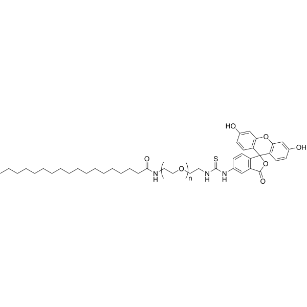
Stearic acid-PEG-FITC, MW 5000 is a PEG lipid which forms micelles in water and can be used for drug delivery applications. The FITC fluorescent can be easily traced by miscroscopy. FITC is a green dye with peak absorption at 494 nm and maximum emission at 520 nm and can be used for staining biological samples or nanoparticles. FITC can be easily traced by fluorescence microscopy.
|
-
-
- HY-W440939
-
|
|
Liposome
|
|
|
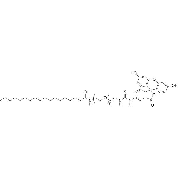
Stearic acid-PEG-FITC, MW 3400 is a PEG lipid which forms micelles in water and can be used for drug delivery applications. The FITC fluorescent can be easily traced by miscroscopy. FITC is a green dye with peak absorption at 494 nm and maximum emission at 520 nm and can be used for staining biological samples or nanoparticles. FITC can be easily traced by fluorescence microscopy.
|
-
-
- HY-151801
-
|
|
mAChR
|
Others
|
|
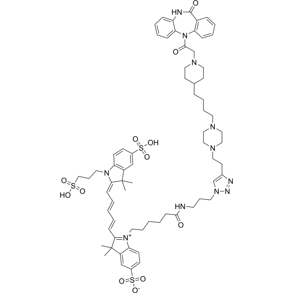
DIBA-Cy5 is a fluorescent DIBA antagonist made up be DIBA-alkyne binding Cyanine5 fluorophores (Cy5) and polyethylene glycol (PEG) biomolecules. DIBA-Cy5 can serve as a fluorescent ligand, suitable for probe attachment through click chemistry. DIBA-Cy5 exerts a high binding affinity to type-2 mAChR (M2R) with the Kd value of 1.80 nM, can directly stain M2R receptors in the sinoatrial node of a mouse heart .
|
-
-
- HY-N6716
-
|
|
Fungal
Antibiotic
|
Infection
|
|
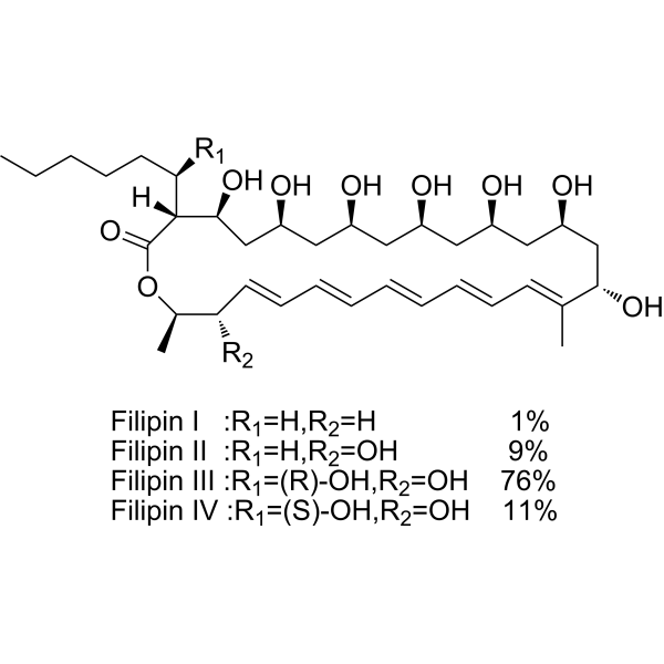
Filipin complex is a potent polyene macrolide antifungal antibiotic. Filipin complex inserts into membranes and sequester cholesterol into complexes and inhibits PRRSV entry. The Filipin complex consists of about 75.8% Filipin III (HY-N6718), 10.8% Filipin IV, 9.1% Filipin II, and 1.2% Filipin I .
|
-
-
- HY-D1346
-
|
|
Fluorescent Dye
|
Others
|
|
610CP is a new type of actin labeling dye. It dissolves in organic solvents. In DMSO the 610CP excitation/emission wavelength is between 609 and 634 nm. 610CP is a fluorescent dye that penetrates living cells. Upon cell entry, 610CP binds to Bromo-des-methyl-Jasplakinolide Therefore, 610CP dye can be used to stain actin fluorescence images with low background and high resolution.
|
-
-
- HY-138658
-
|
JF526, SE; JF526, NHS
|
Fluorescent Dye
|
Others
|
|
Janelia Fluor 526, SE (JF526,SE) is a fluorogenic yellow fluorescent dye that contains NHS ester group. JF526 is a versatile scaffold for fluorogenic ligands, including labels for genetically encoded self-labeling protein tags and stains for endogenous structures . Janelia Fluor products are licensed under U.S. Pat. Nos. 9,933,417, 10,018,624 and 10,161,932 and other patents from Howard Hughes Medical Institute.
|
-
-
- HY-D1246
-
|
|
DNA Stain
|
Others
|
|
Ethidium monoazide bromide is a DNA intercalating fluorescent dye that enters bacteria with damaged membranes. Ethidium monoazide bromide can be covalently linked to DNA by photoactivation. Ethidium monoazide bromide stains only dead cells . Ethidium monoazide (bromide) is a click chemistry reagent, it contains an Azide group and can undergo copper-catalyzed azide-alkyne cycloaddition reaction (CuAAc) with molecules containing Alkyne groups. Strain-promoted alkyne-azide cycloaddition (SPAAC) can also occur with molecules containing DBCO or BCN groups.
|
-
-
- HY-D0814
-
|
4',6-Diamidino-2-phenylindole dihydrochloride
|
DNA Stain
|
Others
|
|
DAPI dihydrochloride is a DAPI dye. DAPI is a fluorescent dye that binds strongly to DNA. It binds to the AT base pair of the double-stranded DNA minor groove, and one DAPI molecule can occupy three base pair positions. The fluorescence intensity of DAPI molecules bound to double-stranded DNA is increased by about 20 times, and it is commonly observed with fluorescence microscopy, and the amount of DNA can be determined based on the intensity of fluorescence. In addition, because DAPI can pass through intact cell membranes, it can be used to stain both live and fixed cells .
|
-
-
- HY-D1068
-
|
DBCO-Sulfo-Cy5
|
Fluorescent Dye
|
Others
|
|
Cy5-DBCO (DBCO-Sulfo-Cy5) is a near-infrared (NIR)red fluorescent dye with λabsand λem of 646nm and 670 nm, respectively. Cy5-DBCO (DBCO-Sulfo-Cy5) is not suitable for staining intracellular components of permeabilezed cell, it may exhibits a high background. Cy5-DBCO is a click chemistry reagent, it contains a DBCO group that can undergo strain-promoted alkyne-azide cycloaddition (SPAAC) with molecules containing Azide groups.
|
-
-
- HY-D0079
-
|
Hydroethidine; PD-MY 003
|
Fluorescent Dye
|
Others
|
|
Dihydroethidium, also known as DHE, is a peroxide indicator. Dihydroethidium penetrates cell membranes to form a fluorescent protein complex with blue fluoresces. After entering the cells, Dihydroethidium is mainly localized in the cell membrane, cytoplasm and nucleus, and the staining effect is the strongest in the nucleus. Dihydroethidium produces inherent blue fluorescence with a maximum excitation wavelength of 370 nm and a maximum emission wavelength of 420 nm; after dehydrogenation, Dihydroethidium combines with RNA or DNA to produce red fluorescence with a maximum excitation wavelength of 300 nm and a maximum emission wavelength of 610 nm. 535 nm can also be used as the excitation wavelength for actual observation .
|
-
-
- HY-D0938
-
CFDA-SE
Maximum Cited Publications
53 Publications Verification
5(6)-Carboxyfluorescein diacetate succinimidyl ester; 5(6)-CFDA N-succinmidyl ester
|
Fluorescent Dye
|
Others
|
|
CFDA-SE is a fluorescent dye that can penetrate the cell membrane. It can react with the free amine group in the cytoskeleton protein inside the cell, and finally form a protein complex with fluorescence. After entering the cell, CFDA-SE locates in the cell membrane, cytoplasm and nucleus, and the fluorescence staining is strongest in the nucleus .
CFDA-SE dye can be uniformly inherited by the cells with cell division and proliferation, and its attenuation is proportional to the number of cell divisions. This phenomenon can be detected and analyzed by flow cytometry under the excitation light of 488 nm, and can be used to detect the proliferation of cells .
|
-
-
- HY-D1804
-
|
|
Fluorescent Dye
|
Others
|
|
Vari Fluor 680-Streptavidin is a dye marker of Vari Fluor-streptavidin consisting of labeling streptavidin with a Vari Fluor series of fluorescent probes. Streptavidin is a high-affinity tetramer protein, each tetramer consisting of four identical streptavidin subunits. Streptavidin binds to biotin specifically via a reversible non-covalent effect. Streptavidin can achieve rapid and efficient detection of biotin markers, and is often used in immunofluorescence (IF), enzyme-linked immunosorbent assay (ELISA), immunohistochemical staining (IFH), in situ hybridization (ISH) and other experiments. Ex/Em=680 nm/701 nm.
|
-

-
- HY-D1805
-
|
|
Fluorescent Dye
|
Others
|
|
Vari Fluor 647-Streptavidin is a dye marker of Vari Fluor-streptavidin consisting of labeling streptavidin with a Vari Fluor series of fluorescent probes. Streptavidin is a high-affinity tetramer protein, each tetramer consisting of four identical streptavidin subunits. Streptavidin binds to biotin specifically via a reversible non-covalent effect. Streptavidin can achieve rapid and efficient detection of biotin markers, and is often used in immunofluorescence (IF), enzyme-linked immunosorbent assay (ELISA), immunohistochemical staining (IFH), in situ hybridization (ISH) and other experiments. Ex/Em=650 nm/665 nm.
|
-

- HY-D1806
-
|
|
Fluorescent Dye
|
Others
|
|
Vari Fluor 594-Streptavidin is a dye marker of Vari Fluor-streptavidin consisting of labeling streptavidin with a Vari Fluor series of fluorescent probes. Streptavidin is a high-affinity tetramer protein, each tetramer consisting of four identical streptavidin subunits. Streptavidin binds to biotin specifically via a reversible non-covalent effect. Streptavidin can achieve rapid and efficient detection of biotin markers, and is often used in immunofluorescence (IF), enzyme-linked immunosorbent assay (ELISA), immunohistochemical staining (IFH), in situ hybridization (ISH) and other experiments. Ex/Em=590 nm/617 nm.
|
-

- HY-D1807
-
|
|
Fluorescent Dye
|
Others
|
|
Vari Fluor 555-Streptavidin is a dye marker of Vari Fluor-streptavidin consisting of labeling streptavidin with a Vari Fluor series of fluorescent probes. Streptavidin is a high-affinity tetramer protein, each tetramer consisting of four identical streptavidin subunits. Streptavidin binds to biotin specifically via a reversible non-covalent effect. Streptavidin can achieve rapid and efficient detection of biotin markers, and is often used in immunofluorescence (IF), enzyme-linked immunosorbent assay (ELISA), immunohistochemical staining (IFH), in situ hybridization (ISH) and other experiments. Ex/Em=555 nm/565 nm.
|
-

- HY-D1808
-
|
|
Fluorescent Dye
|
Others
|
|
Vari Fluor 488-Streptavidin is a dye marker of Vari Fluor-streptavidin consisting of labeling streptavidin with a Vari Fluor series of fluorescent probes. Streptavidin is a high-affinity tetramer protein, each tetramer consisting of four identical streptavidin subunits. Streptavidin binds to biotin specifically via a reversible non-covalent effect. Streptavidin can achieve rapid and efficient detection of biotin markers, and is often used in immunofluorescence (IF), enzyme-linked immunosorbent assay (ELISA), immunohistochemical staining (IFH), in situ hybridization (ISH) and other experiments. Ex/Em=490 nm/515 nm.
|
-

- HY-D1809
-
|
|
Fluorescent Dye
|
Others
|
|
Vari Fluor 405-Streptavidin is a dye marker of Vari Fluor-streptavidin consisting of labeling streptavidin with a Vari Fluor series of fluorescent probes. Streptavidin is a high-affinity tetramer protein, each tetramer consisting of four identical streptavidin subunits. Streptavidin binds to biotin specifically via a reversible non-covalent effect. Streptavidin can achieve rapid and efficient detection of biotin markers, and is often used in immunofluorescence (IF), enzyme-linked immunosorbent assay (ELISA), immunohistochemical staining (IFH), in situ hybridization (ISH) and other experiments. Ex/Em=405 nm/431 nm.
|
-

- HY-118411
-
|
|
PROTAC Linkers
|
Cancer
|
|
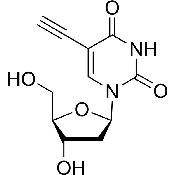
5-Ethynyl-2'-deoxyuridine (EdU), a thymidine analogue, is incorporated into cellular DNA during DNA replication and the subsequent reaction of EdU with a fluorescent azide in a “Click” reaction. EdU staining is a fast, sensitive and reproducible method to study cell proliferation . 5-Ethynyl-2'-deoxyuridine is an alkyl chain-based PROTAC linker that can be used in the synthesis of PROTACs . 5-Ethynyl-2'-deoxyuridine is a click chemistry reagent, it contains an Alkyne group and can undergo copper-catalyzed azide-alkyne cycloaddition (CuAAc) with molecules containing Azide groups.
|
-
- HY-D1396
-
Br-DAPI
3 Publications Verification
|
Fluorescent Dye
DNA Stain
|
Others
|
|
Br-DAPI is a marker dye in DAPI series. DAPI is a fluorescent dye that binds strongly to DNA. It binds to the AT base pair of the double-stranded DNA minor groove, and one DAPI molecule can occupy three base pair positions. The fluorescence intensity of DAPI molecules bound to double-stranded DNA is increased by about 20 times, and it is commonly observed with fluorescence microscopy, and the amount of DNA can be determined based on the intensity of fluorescence. In addition, because DAPI can pass through intact cell membranes, it can be used to stain both live and fixed cells . Storage: Keep away from light.
|
-
- HY-D0952
-
|
|
Parasite
|
Others
|
|
Acridine Orange base is a cell-permeable fluorescent dye that stains organisms (bacteria, parasites, viruses, etc.) bright orange and, when used under appropriate conditions (pH=3.5, Ex=460 nm), distinguishes human cells in green for detection by fluorescence microscopy. Acridine Orange base fluoresces green when bound to dsDNA (Ex=488, Em=520-524) and red when bound to ssDNA (Ex=457, Em=630-644) or ssRNA (Ex=457, Em=630-644), also can be used in cell cycle assays .
|
-
| Cat. No. |
Product Name |
Type |
-
- HY-101888
-
-
- HY-Y0016
-
|
Basic Violet 10; Brilliant Pink B; Rhodamine O; Tetraethylrhodamine
|
Fluorescent Dyes/Probes
|
|
Rhodamine B is a staining fluorescent dye, commonly used for dyeing textiles, paper, soap, leather, and agents.
|
-
- HY-117070
-
|
|
Fluorescent Dyes/Probes
|
|
TO-PRO-3 iodide is a highly efficient blue fluorescent dye that can stain cytoplasm as a cell tracer.
|
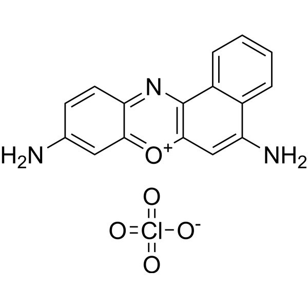
-
- HY-101889
-
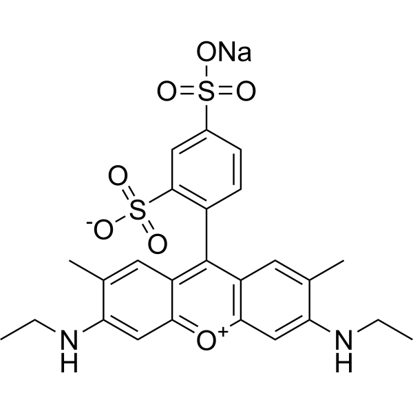
|
Oxazine 9 perchlorate
|
Fluorescent Dyes/Probes
|
|
Cresyl Violet perchlorate is a red fluorescent stain, which can be used to stain neurons.
|
-
- HY-D1775
-
|
|
Fluorescent Dyes/Probes
|
|
Lysotracker blue DND-22 is a blue-fluorescent probe for staining acidic compartments in live cells.
|
-
- HY-D0163
-
|
|
DNA Stain
|
|
Methyl Green is a potent fluorescent dye. Methyl Green is a DNA stains of cells and electrophoretic gels. Methyl Green can be used as a stain for direct measuring of viability by both microscopy and flow cytometry, with peaks at 633 and 677 nm .
|
-
- HY-D0021
-
|
EtBr; Homidium bromide
|
Fluorescent Dyes/Probes
|
|
Ethidium bromide is an intercalating agent commonly used as a fluorescent tag (nucleic acid stain) in molecular biology laboratories for techniques such as agarose gel electrophoresis.
|
-
- HY-D1475
-
|
|
Fluorescent Dyes/Probes
|
|
SIR-6-COOH is a fluorescent dye. SIR-6-COOH can be used for staining proteins in live-cell STED imaging
|
-
- HY-D1385
-
|
|
Fluorescent Dyes/Probes
|
|
JF526–Pepstatin A TFA is a fluorescent dye that can be used for lysosomal staining in live cells. The excitation maximum is 530 nm and the emission maximum is 549 nm .
|
-
- HY-D0718
-
|
Nile Blue A oxazone; Phenoxazone 9
|
Fluorescent Dyes/Probes
|
|
Nile red (Nile blue oxazone) is a lipophilic stain. Nile red has environment-sensitive fluorescence. Nile red is intensely fluorescent in a lipid-rich environment while it has minimal fluorescence in aqueous media. Nile red is an excellent vital stain for the detection of intracellular lipid droplets by fluorescence microscopy and flow cytof uorometry. Nile red stains intracellular lipid droplets red. The fluorescence wavelength is 559/635 nm .
|
-
- HY-D0127
-
|
|
Fluorescent Dyes/Probes
|
|
Merocyanin 540 is a fluorescent membrane probe that selectively stains the membranes of a wide variety of electrically excitable cells, but not those of nonexcitable cells (Ex/Em: 540/580 nm) .
|
-
- HY-D1493
-
|
|
Fluorescent Dyes/Probes
|
|
FIM-1 is a fluorescent PKC (protein kinase C) probe that can be used for mitochondrial staining. FIM-1 inhibits PKC and acts as ATP-competitive catalytic site inhibitor .
|
-
- HY-137896
-
|
|
Fluorescent Dyes/Probes
|
|
4-Acetamido-4'-isothiocyanatostilbene-2,2'-disulfonic acid disodium is a fluorescent dye. 4-Acetamido-4'-isothiocyanatostilbene-2,2'-disulfonic acid disodium can be used to demonstrate retrograde axonal transport to label secondary antibodies and as a fluorescent whole cell stain .
|
-
- HY-D1691
-
|
|
Fluorescent Dyes/Probes
|
|
BODIPY-581/591 NHS ester is a bright, red fluorescent dye (excitation: 581 nm; emission: 591 nm). BODIPY-581/591 NHS ester shows unique hydrophobic properties for staining lipids, membranes, and other lipophilic compounds .
|
-
- HY-D1249
-
|
|
Fluorescent Dyes/Probes
|
|
Calcein mixture of isomers is a calcium-dependent fluorescent molecule. Calcein mixture of isomers can be used to study bone metabolism (in vivo) and to stain depressed areas (in vitro). Calcein mixture of isomers can also be used for fluorometry and EDTA titration of calcium.
|
-
- HY-D1630
-
|
|
Fluorescent Dyes/Probes
|
|
4-Di-10-ASP is a fluorescent lipophilic tracer (Excitation 485 nm; Emission 620 nm). 4-Di-10-ASP can be used to stain phospholipid membranes in a specific manner .
|
-
- HY-D0950A
-
|
|
DNA Stain
|
|
Methyl Green zinc chloride is a potent fluorescent dye. Methyl Green zinc chloride is a DNA stains of cells and electrophoretic gels. Methyl Green zinc chloride can be used as direct measuring of viability by both microscopy and flow cytometry, with peaks at 633 and 677 nm .
|
-
- HY-D1421
-
PKH 67
1 Publications Verification
|
Fluorescent Dyes/Probes
|
|
PKH67 is a fluorescent cell binding dye with green fluorescence. PKH67 can stain the cell membrane and the Ex/Em is 490/502 nm. PKH67 is often used in combination with the non-specific red fluorescent dye PKH26 (Ex/Em=551/567 nm) to label cells, detect cell proliferation in vitro, and trace cells in vitro and in vivo .
|
-
- HY-D1074
-
|
3,3'-Dipropyloxacarbocyanine iodide
|
Fluorescent Dyes/Probes
|
|
DiOC3(3) (3,3'-Dipropyloxacarbocyanine iodide) is a green fluorescent lipophilic dye with cell membrane permeability. DiOC3(3) can be used to stain cell membranes and other lipid-soluble biological structures .
|
-
- HY-D1020
-
|
7-AAD
|
DNA Stain
|
|
7-Aminoactinomycin D (7-AAD) a fluorescent DNA stain, is a potent RNA polymerase inhibitor. 7-Aminoactinomycin D selectively binds to GC regions of the DNA. 7-Aminoactinomycin D also has antibacterial effects .
|
-
- HY-135056
-
|
|
Fluorescent Dyes/Probes
|
|
Mito-Tracker Green is a green fluorescent dye that selectively accumulates in the mitochondrial matrix. MitoTracker Green FM covalently binds mitochondrial proteins by reacting with free mercaptan of cysteine residues, allowing staining of mitochondrial membrane potential independent of membrane potential. Excitation/emission wavelength 490/523 nm.
|
-
- HY-D0093
-
|
EthD-1
|
DNA Stain
|
|
Ethidium homodimer (EthD-1) is a high-affinity fluorescent nucleic acid dye commonly used to stain mammals, bacteria, yeast, and fungi. Ethidium homodimer binds to DNA or RNA, enhancing fluorescence more than 30 times. The Ethidium homodimer has a strong positive charge, so it cannot cross cell membranes and stain living cells; But the Ethidium homodimer can cross the disordered region of the dead cell membrane to reach the nucleus and embed the DNA double strand to produce red fluorescence. Therefore, Ethidium homodimer is a relatively sensitive nucleic acid stain that can accurately detect nucleic acids in solution or in decomposing cells. Ethidium homodimer binds DNA, Ex/Em=528/617 nm .
|
-
- HY-103240
-
|
|
Dyes
|
|
Methoxy-X04 is a fluorescent dye that crosses the blood-brain barrier and selectively binds to beta-pleated sheets found in dense core amyloid Aβ plaques. Methoxy-X04 retains in vitro binding affinity for amyloid b (Ab) fibrils (Ki= 26.8 nM). Methoxy-X04 is fluorescent and stains plaques, tangles, and cerebrovascular amyloid in postmortem sections of AD brain with good specificity .
|
-
- HY-135009
-
|
DASPI
|
Fluorescent Dyes/Probes
|
|
2-Di-1-ASP (DASPI; Compound 18a) is a mono-stryryl dye, and widely used as mitochondrial stain and groove-binding fluorescent probes for double-stranded DNA. 2-Di-1-ASP is selective for G-quadruplex (G4) and double-stranded DNA .
|
-
- HY-W094758A
-
|
|
Fluorescent Dyes/Probes
|
|
4-Di-1-ASP is a styryl dye used to stain glioma cells in living brain tissue for analysis of cell structure, viability, proliferation and endocytosis, cytokinesis and phagocytosis, as well as for observation of mitochondrial structures in living cells. 4-Di-1-ASP fluoresces green when imaged microscopically (λex /λem = 475/606 nm) .
|
-
- HY-D1738
-
|
4',6-Diamidino-2-phenylindole dilactate
|
Fluorescent Dyes/Probes
|
|
DAPI (dilactate) is a blue fluorescent dye that preferentially binds dsDNA and binds to minor groove AT clusters. DAPI (dilactate) is combined with dsDNA, and the fluorescence was enhanced about 20-fold. DAPI (dilactate) can be used to identify the cell cycle and specifically stains the nucleus but not the cytoplasm. DAPI (dilactate) form is more soluble in water than DAPI (dihydrochloride) form.
|
-
- HY-151801
-
|
|
Fluorescent Dyes/Probes
|
|
DIBA-Cy5 is a fluorescent DIBA antagonist made up be DIBA-alkyne binding Cyanine5 fluorophores (Cy5) and polyethylene glycol (PEG) biomolecules. DIBA-Cy5 can serve as a fluorescent ligand, suitable for probe attachment through click chemistry. DIBA-Cy5 exerts a high binding affinity to type-2 mAChR (M2R) with the Kd value of 1.80 nM, can directly stain M2R receptors in the sinoatrial node of a mouse heart .
|
-
- HY-N6716
-
|
|
Fluorescent Dyes/Probes
|
|
Filipin complex is a potent polyene macrolide antifungal antibiotic. Filipin complex inserts into membranes and sequester cholesterol into complexes and inhibits PRRSV entry. The Filipin complex consists of about 75.8% Filipin III (HY-N6718), 10.8% Filipin IV, 9.1% Filipin II, and 1.2% Filipin I .
|
-
- HY-D1346
-
|
|
Fluorescent Dyes/Probes
|
|
610CP is a new type of actin labeling dye. It dissolves in organic solvents. In DMSO the 610CP excitation/emission wavelength is between 609 and 634 nm. 610CP is a fluorescent dye that penetrates living cells. Upon cell entry, 610CP binds to Bromo-des-methyl-Jasplakinolide Therefore, 610CP dye can be used to stain actin fluorescence images with low background and high resolution.
|
-
- HY-138658
-
|
JF526, SE; JF526, NHS
|
Fluorescent Dyes/Probes
|
|
Janelia Fluor 526, SE (JF526,SE) is a fluorogenic yellow fluorescent dye that contains NHS ester group. JF526 is a versatile scaffold for fluorogenic ligands, including labels for genetically encoded self-labeling protein tags and stains for endogenous structures . Janelia Fluor products are licensed under U.S. Pat. Nos. 9,933,417, 10,018,624 and 10,161,932 and other patents from Howard Hughes Medical Institute.
|
-
- HY-D1246
-
|
|
DNA Stain
|
|
Ethidium monoazide bromide is a DNA intercalating fluorescent dye that enters bacteria with damaged membranes. Ethidium monoazide bromide can be covalently linked to DNA by photoactivation. Ethidium monoazide bromide stains only dead cells . Ethidium monoazide (bromide) is a click chemistry reagent, it contains an Azide group and can undergo copper-catalyzed azide-alkyne cycloaddition reaction (CuAAc) with molecules containing Alkyne groups. Strain-promoted alkyne-azide cycloaddition (SPAAC) can also occur with molecules containing DBCO or BCN groups.
|
-
- HY-D0814
-
|
4',6-Diamidino-2-phenylindole dihydrochloride
|
Fluorescent Dyes/Probes
|
|
DAPI dihydrochloride is a DAPI dye. DAPI is a fluorescent dye that binds strongly to DNA. It binds to the AT base pair of the double-stranded DNA minor groove, and one DAPI molecule can occupy three base pair positions. The fluorescence intensity of DAPI molecules bound to double-stranded DNA is increased by about 20 times, and it is commonly observed with fluorescence microscopy, and the amount of DNA can be determined based on the intensity of fluorescence. In addition, because DAPI can pass through intact cell membranes, it can be used to stain both live and fixed cells .
|
-
- HY-D1068
-
|
DBCO-Sulfo-Cy5
|
Fluorescent Dyes/Probes
|
|
Cy5-DBCO (DBCO-Sulfo-Cy5) is a near-infrared (NIR)red fluorescent dye with λabsand λem of 646nm and 670 nm, respectively. Cy5-DBCO (DBCO-Sulfo-Cy5) is not suitable for staining intracellular components of permeabilezed cell, it may exhibits a high background. Cy5-DBCO is a click chemistry reagent, it contains a DBCO group that can undergo strain-promoted alkyne-azide cycloaddition (SPAAC) with molecules containing Azide groups.
|
-
- HY-D0079
-
|
Hydroethidine; PD-MY 003
|
Fluorescent Dyes/Probes
|
|
Dihydroethidium, also known as DHE, is a peroxide indicator. Dihydroethidium penetrates cell membranes to form a fluorescent protein complex with blue fluoresces. After entering the cells, Dihydroethidium is mainly localized in the cell membrane, cytoplasm and nucleus, and the staining effect is the strongest in the nucleus. Dihydroethidium produces inherent blue fluorescence with a maximum excitation wavelength of 370 nm and a maximum emission wavelength of 420 nm; after dehydrogenation, Dihydroethidium combines with RNA or DNA to produce red fluorescence with a maximum excitation wavelength of 300 nm and a maximum emission wavelength of 610 nm. 535 nm can also be used as the excitation wavelength for actual observation .
|
-
- HY-D0938
-
CFDA-SE
Maximum Cited Publications
53 Publications Verification
5(6)-Carboxyfluorescein diacetate succinimidyl ester; 5(6)-CFDA N-succinmidyl ester
|
Fluorescent Dyes/Probes
|
|
CFDA-SE is a fluorescent dye that can penetrate the cell membrane. It can react with the free amine group in the cytoskeleton protein inside the cell, and finally form a protein complex with fluorescence. After entering the cell, CFDA-SE locates in the cell membrane, cytoplasm and nucleus, and the fluorescence staining is strongest in the nucleus .
CFDA-SE dye can be uniformly inherited by the cells with cell division and proliferation, and its attenuation is proportional to the number of cell divisions. This phenomenon can be detected and analyzed by flow cytometry under the excitation light of 488 nm, and can be used to detect the proliferation of cells .
|
-
- HY-D1804
-
|
|
Dyes
|
|
Vari Fluor 680-Streptavidin is a dye marker of Vari Fluor-streptavidin consisting of labeling streptavidin with a Vari Fluor series of fluorescent probes. Streptavidin is a high-affinity tetramer protein, each tetramer consisting of four identical streptavidin subunits. Streptavidin binds to biotin specifically via a reversible non-covalent effect. Streptavidin can achieve rapid and efficient detection of biotin markers, and is often used in immunofluorescence (IF), enzyme-linked immunosorbent assay (ELISA), immunohistochemical staining (IFH), in situ hybridization (ISH) and other experiments. Ex/Em=680 nm/701 nm.
|
-
- HY-D1805
-
|
|
Dyes
|
|
Vari Fluor 647-Streptavidin is a dye marker of Vari Fluor-streptavidin consisting of labeling streptavidin with a Vari Fluor series of fluorescent probes. Streptavidin is a high-affinity tetramer protein, each tetramer consisting of four identical streptavidin subunits. Streptavidin binds to biotin specifically via a reversible non-covalent effect. Streptavidin can achieve rapid and efficient detection of biotin markers, and is often used in immunofluorescence (IF), enzyme-linked immunosorbent assay (ELISA), immunohistochemical staining (IFH), in situ hybridization (ISH) and other experiments. Ex/Em=650 nm/665 nm.
|
-
- HY-D1806
-
|
|
Dyes
|
|
Vari Fluor 594-Streptavidin is a dye marker of Vari Fluor-streptavidin consisting of labeling streptavidin with a Vari Fluor series of fluorescent probes. Streptavidin is a high-affinity tetramer protein, each tetramer consisting of four identical streptavidin subunits. Streptavidin binds to biotin specifically via a reversible non-covalent effect. Streptavidin can achieve rapid and efficient detection of biotin markers, and is often used in immunofluorescence (IF), enzyme-linked immunosorbent assay (ELISA), immunohistochemical staining (IFH), in situ hybridization (ISH) and other experiments. Ex/Em=590 nm/617 nm.
|
-
- HY-D1807
-
|
|
Dyes
|
|
Vari Fluor 555-Streptavidin is a dye marker of Vari Fluor-streptavidin consisting of labeling streptavidin with a Vari Fluor series of fluorescent probes. Streptavidin is a high-affinity tetramer protein, each tetramer consisting of four identical streptavidin subunits. Streptavidin binds to biotin specifically via a reversible non-covalent effect. Streptavidin can achieve rapid and efficient detection of biotin markers, and is often used in immunofluorescence (IF), enzyme-linked immunosorbent assay (ELISA), immunohistochemical staining (IFH), in situ hybridization (ISH) and other experiments. Ex/Em=555 nm/565 nm.
|
-
- HY-D1808
-
|
|
Dyes
|
|
Vari Fluor 488-Streptavidin is a dye marker of Vari Fluor-streptavidin consisting of labeling streptavidin with a Vari Fluor series of fluorescent probes. Streptavidin is a high-affinity tetramer protein, each tetramer consisting of four identical streptavidin subunits. Streptavidin binds to biotin specifically via a reversible non-covalent effect. Streptavidin can achieve rapid and efficient detection of biotin markers, and is often used in immunofluorescence (IF), enzyme-linked immunosorbent assay (ELISA), immunohistochemical staining (IFH), in situ hybridization (ISH) and other experiments. Ex/Em=490 nm/515 nm.
|
-
- HY-D1809
-
|
|
Dyes
|
|
Vari Fluor 405-Streptavidin is a dye marker of Vari Fluor-streptavidin consisting of labeling streptavidin with a Vari Fluor series of fluorescent probes. Streptavidin is a high-affinity tetramer protein, each tetramer consisting of four identical streptavidin subunits. Streptavidin binds to biotin specifically via a reversible non-covalent effect. Streptavidin can achieve rapid and efficient detection of biotin markers, and is often used in immunofluorescence (IF), enzyme-linked immunosorbent assay (ELISA), immunohistochemical staining (IFH), in situ hybridization (ISH) and other experiments. Ex/Em=405 nm/431 nm.
|
-
- HY-D0952
-
|
|
Fluorescent Dyes/Probes
|
|
Acridine Orange base is a cell-permeable fluorescent dye that stains organisms (bacteria, parasites, viruses, etc.) bright orange and, when used under appropriate conditions (pH=3.5, Ex=460 nm), distinguishes human cells in green for detection by fluorescence microscopy. Acridine Orange base fluoresces green when bound to dsDNA (Ex=488, Em=520-524) and red when bound to ssDNA (Ex=457, Em=630-644) or ssRNA (Ex=457, Em=630-644), also can be used in cell cycle assays .
|
| Cat. No. |
Product Name |
Type |
-
- HY-D1021
-
-
- HY-107629
-
-
- HY-W440940
-
|
|
Drug Delivery
|
|
Stearic acid-PEG-FITC, MW 5000 is a PEG lipid which forms micelles in water and can be used for drug delivery applications. The FITC fluorescent can be easily traced by miscroscopy. FITC is a green dye with peak absorption at 494 nm and maximum emission at 520 nm and can be used for staining biological samples or nanoparticles. FITC can be easily traced by fluorescence microscopy.
|
-
- HY-W440939
-
|
|
Drug Delivery
|
|
Stearic acid-PEG-FITC, MW 3400 is a PEG lipid which forms micelles in water and can be used for drug delivery applications. The FITC fluorescent can be easily traced by miscroscopy. FITC is a green dye with peak absorption at 494 nm and maximum emission at 520 nm and can be used for staining biological samples or nanoparticles. FITC can be easily traced by fluorescence microscopy.
|
| Cat. No. |
Product Name |
Category |
Target |
Chemical Structure |
| Cat. No. |
Product Name |
|
Classification |
-
- HY-D1068
-
|
DBCO-Sulfo-Cy5
|
|
DBCO
Labeling and Fluorescence Imaging
|
|
Cy5-DBCO (DBCO-Sulfo-Cy5) is a near-infrared (NIR)red fluorescent dye with λabsand λem of 646nm and 670 nm, respectively. Cy5-DBCO (DBCO-Sulfo-Cy5) is not suitable for staining intracellular components of permeabilezed cell, it may exhibits a high background. Cy5-DBCO is a click chemistry reagent, it contains a DBCO group that can undergo strain-promoted alkyne-azide cycloaddition (SPAAC) with molecules containing Azide groups.
|
-
- HY-118411
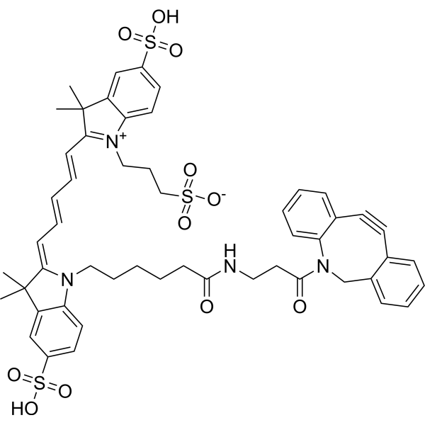
-
|
|
|
PROTAC Synthesis
Alkynes
|
|
5-Ethynyl-2'-deoxyuridine (EdU), a thymidine analogue, is incorporated into cellular DNA during DNA replication and the subsequent reaction of EdU with a fluorescent azide in a “Click” reaction. EdU staining is a fast, sensitive and reproducible method to study cell proliferation . 5-Ethynyl-2'-deoxyuridine is an alkyl chain-based PROTAC linker that can be used in the synthesis of PROTACs . 5-Ethynyl-2'-deoxyuridine is a click chemistry reagent, it contains an Alkyne group and can undergo copper-catalyzed azide-alkyne cycloaddition (CuAAc) with molecules containing Azide groups.
|
-
- HY-D1246
-
|
|
|
Azide
|
|
Ethidium monoazide bromide is a DNA intercalating fluorescent dye that enters bacteria with damaged membranes. Ethidium monoazide bromide can be covalently linked to DNA by photoactivation. Ethidium monoazide bromide stains only dead cells . Ethidium monoazide (bromide) is a click chemistry reagent, it contains an Azide group and can undergo copper-catalyzed azide-alkyne cycloaddition reaction (CuAAc) with molecules containing Alkyne groups. Strain-promoted alkyne-azide cycloaddition (SPAAC) can also occur with molecules containing DBCO or BCN groups.
|
Your information is safe with us. * Required Fields.
Inquiry Information
- Product Name:
- Cat. No.:
- Quantity:
- MCE Japan Authorized Agent: